






 Back
BackApplications, News, Education
MENISCUS REPAIR: A TISSUE-PRESERVING APPROACH – ARTHROSCOPIC TECHNIQUES – REHABILITATION
Meniscus Repair: A Tissue-Preserving Approach – Arthroscopic Techniques – Rehabilitation
Sửa Chữa Sụn Chêm: Tư Duy Bảo Tồn – Kỹ Thuật Nội Soi – Phục Hồi Chức Năng
English Version | Bản tiếng Việt bên dưới.
1. Fundamental principle: “Meniscus preservation” is the priority
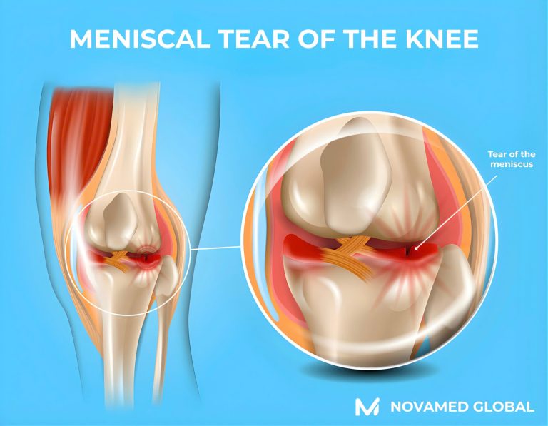
The meniscus acts as a “shock absorber” of the knee joint, helping distribute load and protect the articular cartilage. When the remaining meniscal tissue is insufficient, the risk of osteoarthritis increases significantly. Therefore, modern treatment emphasizes maximal preservation of healthy meniscal tissue.
However, not all meniscal tears require surgery. Operative treatment is considered when conservative management fails or when the tear causes mechanical symptoms such as catching, clicking, or knee locking, particularly in large traumatic tears.
2. The key factor in deciding “repair or resection”: vascular zones (red zone – white zone)
HSS describes the meniscus as having:
- Red zone (peripheral vascular region) approximately 25% → Healing potential is higher and suitable for meniscal repair.
- White zone (inner avascular region) approximately 75% → Healing potential is poor, and repair is more likely to fail. Partial meniscectomy (trimming) is often preferred.
Clinical implication: the same tear pattern but located in different regions (peripheral versus central) may lead to different surgical strategies.
3. Three main treatment strategies for meniscus repair
A. Conservative management (nonoperative treatment)
Applicable to many cases of small tears, degenerative tears, or tears without clear mechanical symptoms:
- Physical therapy with strengthening of the quadriceps and hip musculature and improvement of knee joint kinetic control.
- Activity modification with avoidance of excessive twisting movements.
- Corticosteroid injection may be considered in selected cases to reduce inflammation and pain. It does not repair the tear but may help relieve symptoms.
Goal: pain reduction, functional improvement, and avoidance of unnecessary surgical intervention.
B. Partial meniscectomy (partial removal or trimming)
This procedure removes the irreparable torn portion while preserving as much healthy meniscal tissue as possible. It is commonly performed arthroscopically.
Typical indications:
- Tears located in the white zone with poor vascular supply or meniscal tissue that is frayed or degenerative and unsuitable for repair.
- Complex tears that cannot restore normal shape or stability.
Advantages:
- Recovery is generally faster than meniscal repair. Most patients can bear weight early and return to sports in approximately 4 to 8 weeks, depending on the activity and severity of the tear.
Important counseling points:
- Meniscectomy may relieve mechanical symptoms quickly. However, excessive tissue removal may reduce the protective cushioning function of the meniscus over time. Therefore, the principle is to remove only the minimal necessary amount.
C. Meniscus repair as a tissue preservation strategy
Meniscal repair aims to preserve the meniscal tissue, optimize knee joint biomechanics, and reduce the long-term risk of degeneration.
Typical indications:
- Tears in the red zone where vascular supply is present.
- Vertical longitudinal tears or bucket handle tears are suitable for repair.
- Young and active patients with good-quality meniscal tissue.
Limitations:
- Tears located deep within the white zone or severely degenerative tissue carry a higher risk of nonhealing.
Recovery:
- Crutches and knee bracing are often required in the early phase. Running is usually permitted around 3 to 4 months, and return to sports typically occurs at 6 to 9 months, depending on the repair technique and associated injuries.
4. Meniscus repair techniques: inside out, outside in, all inside
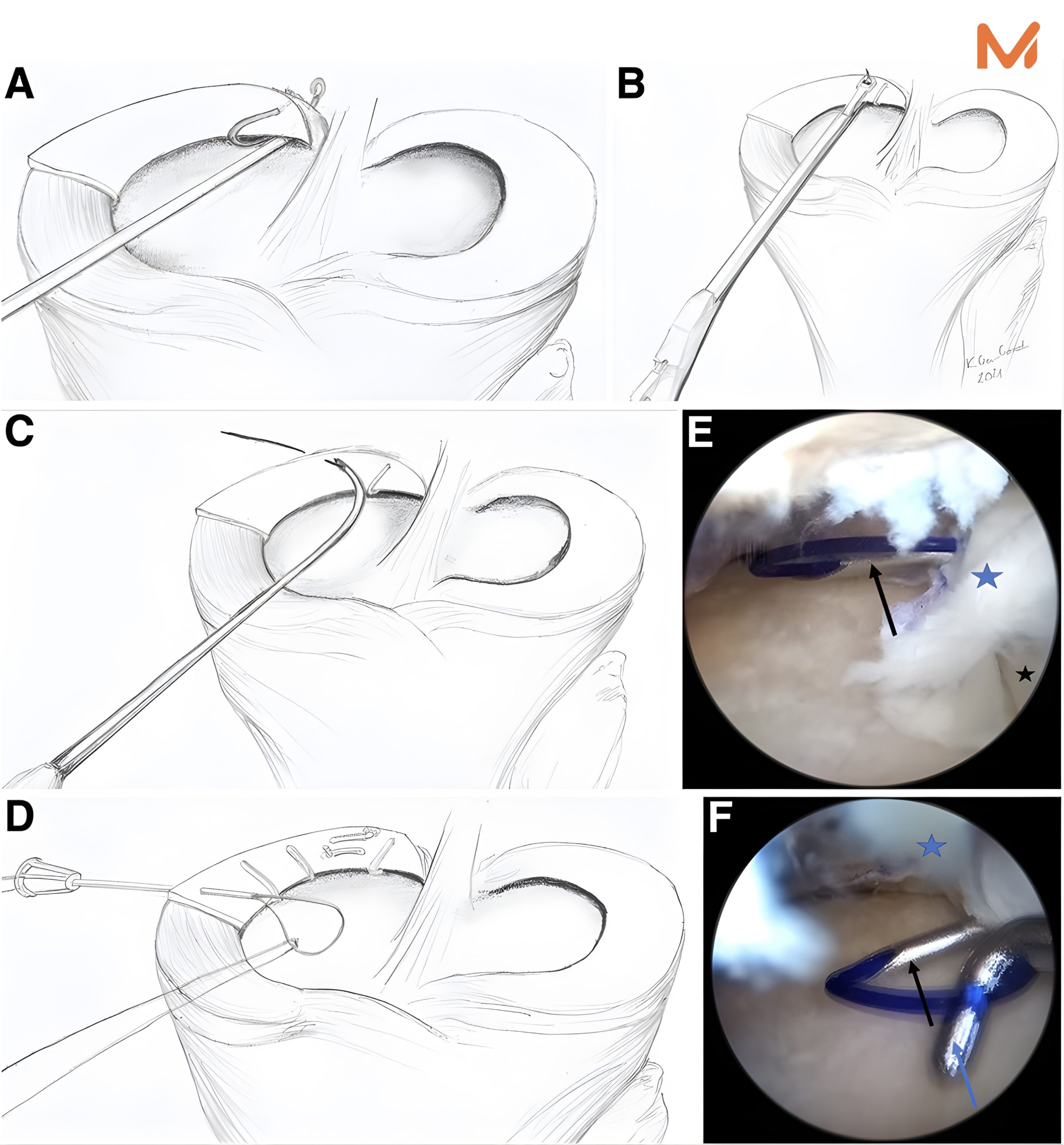
In practice, the choice of technique depends on tear location (anterior, body, posterior), tear morphology, tear length, tissue quality, and the desired postoperative stability.
4.1 All inside repair
- This technique uses suture devices or implants placed arthroscopically, usually without the need for additional large incisions.
- Systematic evidence suggests that functional outcomes, healing rates, and complication rates are comparable to inside-out repair for certain tear patterns, particularly vertical longitudinal and bucket handle tears. Benefits may include reduced operative time and more convenient recovery.
4.2 Inside-out repair
- This is the traditional gold standard for many years, located in the meniscal body and posterior horn, allowing placement of multiple strong sutures.
- An additional incision is usually required to tie the sutures safely and to protect the posterior neurovascular structures. HSS also notes that larger tears may require a supplementary incision to allow for a more robust repair.
4.3 Outside-in repair
- This technique is often useful for tears located in the anterior horn, where needle trajectory and manipulation are more favorable.
- Modern practice frequently uses a hybrid technical approach, selecting suture configurations and devices according to the specific tear location to maximize stability and minimize complications.
5. Special tear patterns requiring specific repair strategies
Beyond typical meniscal body tears, several tear patterns have major biomechanical significance and require specialized repair strategies.
5.1 Meniscal root tears
Root tears, particularly posterior root tears, disrupt the hoop stress mechanism and cause the meniscus to lose its load-distributing function. Many cases are therefore recommended for root repair, often using transtibial pullout techniques or suture anchors. Recent studies suggest that transtibial pullout repair may improve postoperative functional outcomes.
5.2 Ramp lesions and posteromedial meniscus tears
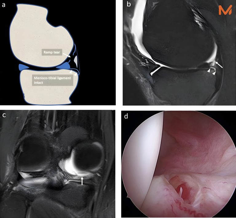
Ramp tears are commonly associated with ACL injury and may contribute to knee instability if missed. Inside-out or all inside techniques may be used depending on the tear configuration.
5.3 Radial tears
Radial tears disrupt the circumferential fibers of the meniscus and significantly affect load transmission. Current trends favor repair when tissue quality permits, using inside-out or all-inside techniques or tunnel-based techniques, depending on tear location and size.
6. When the meniscus cannot be preserved: meniscus allograft transplantation (MAT)
When the injury is too severe and little or no functional meniscal tissue remains, especially in young patients without advanced osteoarthritis, meniscus transplantation from a donor may be considered.
HSS describes the procedure as the removal of the remaining damaged meniscus followed by the placement of the donor graft and fixation with sutures. The early postoperative phase typically requires restricted weight bearing and structured rehabilitation.
The AAOS also emphasizes that MAT may be an option for young and active patients with near-complete meniscal loss in order to reduce pain and protect the joint.
7. Summary of treatment selection based on a decision framework
- Peripheral tears in the red zone with good tissue quality and favorable tear patterns → Meniscal repair is preferred.
- Central tears in the white zone with degenerative or complex features → Selective partial meniscectomy is usually performed.
- Biomechanically significant tear patterns such as root, ramp, and radial tears → Specialized repair techniques should be considered to restore meniscal function.
- Severe meniscal deficiency in young patients without advanced osteoarthritis → Meniscus transplantation may be considered.
8. Practical advice for physicians and patients
- The decision between repair and resection should not be made empirically. It depends on vascular supply and tissue quality.
- Meniscectomy should follow the principle of minimal necessary resection because greater meniscal loss increases the risk of joint degeneration.
- Meniscal repair requires a longer rehabilitation period but aims to preserve long-term joint health.
- Biomechanically disruptive meniscal tears, including root, radial, and ramp lesions, may severely compromise meniscal function and should not be considered simple meniscal tears.
1. Nguyên tắc nền tảng: “Bảo tồn sụn chêm” là ưu tiên
Sụn chêm là “bộ giảm xóc” của khớp gối, giúp phân bố lực và bảo vệ sụn khớp. Khi lượng sụn chêm còn lại quá ít, nguy cơ thoái hóa khớp tăng rõ rệt. Vì vậy, điều trị hiện đại đặt mục tiêu bảo tồn tối đa mô sụn chêm lành.
Tuy nhiên, không phải mọi rách sụn chêm đều cần mổ. Phẫu thuật được cân nhắc khi điều trị bảo tồn thất bại hoặc rách gây triệu chứng cơ học (kẹt, lục khục, “khóa” gối), đặc biệt là rách lớn do chấn thương.
2. Mấu chốt quyết định “khâu hay cắt”: vùng tưới máu (red zone – white zone)
HSS mô tả meniscus có:
- “Red zone” (vùng ngoại vi có tưới máu) ~ 25% → Có khả năng lành, phù hợp để khâu sửa.
- “White zone” (vùng trong không tưới máu) ~ 75% → Khả năng lành kém, khâu dễ thất bại → Thường chọn cắt bán phần (trim/partial meniscectomy).
Ý nghĩa lâm sàng: cùng một “vết rách” nhưng khác vị trí (ngoại vi vs trung tâm) sẽ dẫn tới lựa chọn phẫu thuật khác nhau.
3. Ba nhóm chiến lược điều trị “sửa chữa sụn chêm”
A. Điều trị bảo tồn (không phẫu thuật)
Áp dụng cho nhiều trường hợp rách nhỏ, rách thoái hóa, triệu chứng không cơ học rõ:
- Vật lý trị liệu, tăng sức mạnh cơ đùi-hông, cải thiện kiểm soát động học khớp gối
- Điều chỉnh hoạt động, giảm các động tác xoay vặn
- Tiêm corticosteroid trong một số trường hợp chọn lọc để giảm viêm/đau (không “vá” vết rách nhưng hỗ trợ triệu chứng)
Mục tiêu: giảm đau, cải thiện chức năng, tránh can thiệp không cần thiết.
B. Cắt sụn chêm bán phần (Partial meniscectomy / “trimming”)
Đây là thủ thuật cắt bỏ phần rách không còn khả năng bảo tồn, giữ lại phần mô lành tối đa; thường được thực hiện qua nội soi.
Chỉ định điển hình:
- Rách ở white zone (kém tưới máu) hoặc mô sụn chêm “tơi”/thoái hóa, không đủ chất lượng để khâu
- Rách phức tạp không tái tạo được hình dạng/độ vững
Ưu điểm:
- Hồi phục nhanh hơn khâu sụn chêm: đa số có thể chịu lực sớm, trở lại thể thao khoảng 4-8 tuần tùy môn và mức độ.
Điểm cần tư vấn rõ:
- Meniscectomy giúp hết triệu chứng cơ học nhanh, nhưng về dài hạn có thể làm giảm “đệm” bảo vệ sụn khớp nếu lấy mô nhiều. Vì vậy, nguyên tắc là cắt tối thiểu cần thiết.
C. Khâu sửa sụn chêm – “điều trị bảo tồn mô”
Khâu sụn chêm nhằm giữ lại mô meniscus, tối ưu sinh cơ học khớp gối và giảm nguy cơ thoái hóa về lâu dài.
Chỉ định điển hình:
- Rách ở red zone (ngoại vi có tưới máu)
- Rách dọc-dài (vertical longitudinal) / bucket-handle phù hợp khâu
- Bệnh nhân trẻ, hoạt động thể thao; mô sụn chêm còn tốt
Hạn chế:
- Rách sâu trong white zone hoặc mô thoái hóa nặng → Nguy cơ không lành cao
Hồi phục:
- Thường cần nạng và nẹp gối giai đoạn đầu; trở lại chạy khoảng 3-4 tháng, thể thao 6-9 tháng tùy kiểu khâu và tổn thương kèm theo.
4. Các kỹ thuật khâu sụn chêm: inside-out, outside-in, all-inside
Trong thực hành, lựa chọn kỹ thuật dựa trên vị trí rách (trước-giữa-sau), hình thái rách, độ dài vết rách, chất lượng mô, và mục tiêu “độ vững” sau khâu.
4.1. All-inside repair (khâu hoàn toàn nội soi)
- Dùng các “suture device/implant” đặt qua nội soi, thường không cần thêm đường rạch lớn.
- Bằng chứng tổng quan cho thấy kết quả chức năng, tỷ lệ lành và biến chứng tương đương với kỹ thuật inside-out đối với một số dạng rách (đặc biệt là rách dọc–dài và bucket-handle), với lợi ích như giảm thời gian mổ và thuận lợi hồi phục.
4.2. Inside-out repair (khâu từ trong ra ngoài)
- “Chuẩn vàng” truyền thống cho nhiều rách vùng thân–sừng sau, cho phép đặt nhiều mũi khâu chắc.
- Thường cần đường rạch phụ để buộc nút an toàn và bảo vệ cấu trúc mạch thần kinh phía sau. HSS cũng đề cập rằng rách lớn thường cần incision bổ sung để có khâu “robust” hơn.
4.3. Outside-in repair (khâu từ ngoài vào trong)
- Hay hữu ích cho các rách vùng sừng trước/anterior horn, nơi đường đi của kim và thao tác thuận lợi hơn.
- Thực hành hiện đại thường là “hệ kỹ thuật lai”: chọn cấu hình mũi khâu/thiết bị theo từng vùng rách để tối đa độ vững và giảm nguy cơ biến chứng.
5. Những “dạng rách đặc biệt” cần kỹ thuật sửa riêng
Ngoài rách thân meniscus thông thường, một số dạng rách có ý nghĩa sinh cơ học lớn và cần chiến lược sửa khác:
5.1. Rách chân rễ sụn chêm
Root tear (đặc biệt posterior root) làm mất chức năng “vòng đai” (hoop stress) → meniscus gần như mất tác dụng phân bố lực. Vì vậy, nhiều trường hợp được khuyến cáo khâu phục hồi root (thường qua đường hầm xương chày/pull-out hoặc neo). Các nghiên cứu gần đây cho thấy kỹ thuật transtibial pullout có thể cải thiện điểm chức năng sau mổ.
5.2. Ramp lesion và rách vùng sau trong
Ramp tear (thường đi kèm ACL) có thể gây mất vững khớp gối nếu bỏ sót; các kỹ thuật inside-out hoặc all-inside thường được dùng tùy cấu hình rách.
5.3. Rách xuyên tâm
Radial tear “cắt ngang” sợi vòng của meniscus, ảnh hưởng lớn đến tải lực; xu hướng hiện nay là ưu tiên sửa (khi mô cho phép) bằng inside-out/all-inside hoặc kỹ thuật qua đường hầm tùy vị trí và độ rộng.
6. Khi sụn chêm “không còn để giữ”: Ghép sụn chêm (Meniscus allograft transplantation – MAT)
Khi tổn thương quá nặng, gần như không còn meniscus khả dụng (đặc biệt ở bệnh nhân trẻ, chưa thoái hóa khớp nặng), có thể cân nhắc ghép sụn chêm từ người hiến. HSS mô tả: phẫu thuật thường bao gồm cắt bỏ toàn bộ phần còn lại và đặt meniscus ghép, sau đó khâu cố định; giai đoạn đầu cần không chịu lực và phục hồi chức năng theo lộ trình.
AAOS cũng nhấn mạnh MAT là lựa chọn cho bệnh nhân trẻ/hoạt động khi meniscus gần như mất, nhằm giảm đau và bảo vệ khớp.
7. Tóm tắt lựa chọn điều trị theo “bản đồ quyết định”
- Rách ngoại vi (red zone), mô tốt, rách dạng phù hợp → ưu tiên khâu sửa.
- Rách trung tâm (white zone), thoái hóa/phức tạp → Thường cắt bán phần có chọn lọc.
- Root / ramp / radial quan trọng về sinh cơ học → Cân nhắc kỹ thuật sửa chuyên biệt để phục hồi chức năng của meniscus.
- Thiếu meniscus nặng ở bệnh nhân trẻ, khớp chưa thoái hóa nặng → Cân nhắc ghép sụn chêm.
8. Lời khuyên cho bác sĩ & bệnh nhân
- “Khâu hay cắt” không quyết định bằng cảm tính, mà phụ thuộc vào vùng tưới máu và chất lượng mô.
- Meniscectomy nên là “cắt tối thiểu cần thiết”, vì meniscus càng mất nhiều, khớp càng tăng nguy cơ thoái hóa.
- Khâu sụn chêm cần phục hồi dài hơn nhưng mục tiêu là bảo vệ khớp lâu dài.
- Nhóm rách sụn chêm làm mất chức năng cơ học (root, radial, ramp) là các tổn thương có thể “đánh sập” chức năng của meniscus, không nên xem như rách thông thường.