





 Back
BackApplications, News, Education
ARTICULAR CARTILAGE LESIONS OF THE ADULT KNEE: CLASSIFICATION, DIAGNOSIS, AND TREATMENT STRATEGIES FROM CONSERVATIVE MANAGEMENT TO REGENERATION
Articular Cartilage Lesions of the Adult Knee: Classification, Diagnosis, and Treatment Strategies from Conservative Management to Regeneration
Tổn Thương Sụn Khớp Gối Ở Người Trưởng Thành: Phân Loại, Chẩn Đoán Và Các Phương Pháp Điều Trị Từ Bảo Tồn Đến Tái Tạo
English Version | Bản tiếng Việt bên dưới.


Why Are Articular Cartilage Lesions a Unique Challenge?
Articular cartilage is a specialized tissue that covers the surface of bones within joints, allowing smooth articulation and load distribution. However, it is also one of the most difficult tissues in the human body to heal.
Knee articular cartilage is highly specialized hyaline connective tissue, composed of chondrocytes embedded in an extracellular matrix consisting of approximately 90% water, along with proteoglycans, glycosaminoglycans, and predominantly type II collagen. It is organized into four distinct zones, each with a different cell density, matrix composition, and collagen fiber orientation. Its avascular nature creates intrinsic limitations in its capacity for self-repair.
Focal cartilage lesions refer to localized defects or damage to the articular cartilage surface, commonly resulting from trauma, cumulative wear, or underlying joint pathology. If left untreated, these lesions may lead to pain, functional impairment, and progression to osteoarthritis.
Classification of Knee Articular Cartilage Lesions
Classification by depth (ICRS grading system)

Classification by lesion characteristics
Treatment strategies are generally categorized into:
- Palliative (chondroplasty, debridement)
- Reparative (drilling, microfracture)
- Restorative (autologous chondrocyte implantation, osteochondral autograft, osteochondral allograft)
Clinical Presentation and Diagnosis
Clinical features
Articular cartilage lesions can significantly impact daily activities, presenting with:
- Joint swelling
- Pain-related limitation of motion
- Localized pain and joint discomfort
Typical symptoms include:
- Localized knee pain, aggravated by weight-bearing
- Recurrent joint effusion
- Mechanical symptoms (clicking, crepitus)
- Stiffness after prolonged rest
- Reduced range of motion in advanced cases
Imaging
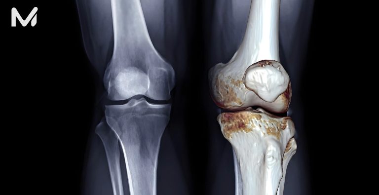
- X-ray: Evaluation of joint space narrowing and associated osteoarthritis
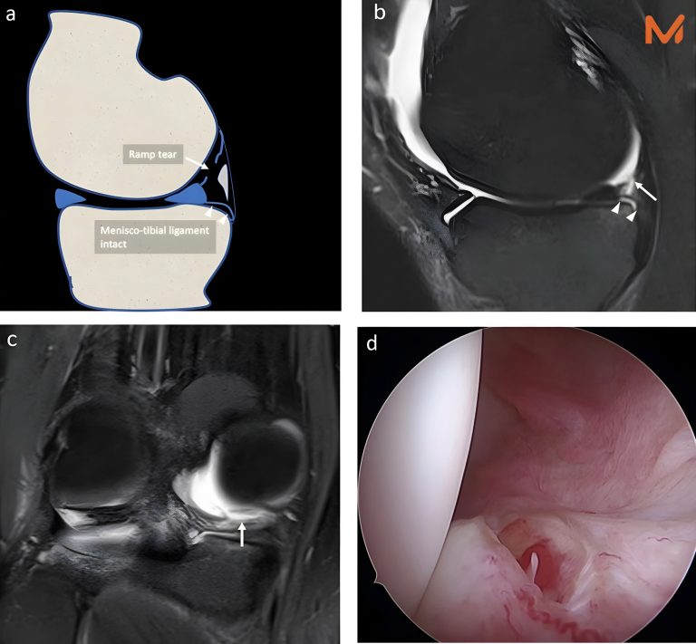
- MRI: Gold standard for assessing lesion location, size, depth, and subchondral bone status
- Arthroscopy: Direct visualization and simultaneous therapeutic intervention
Comprehensive Treatment Spectrum – From Conservative to Restorative
GROUP 1: Conservative and Biological Treatments
Physical therapy and weight management
The foundation of all treatment strategies is aimed at strengthening periarticular muscles and reducing joint loading.
Hyaluronic Acid Injection (Viscosupplementation)
Provides symptomatic relief in knee osteoarthritis. Suitable for mild-to-moderate lesions, particularly in older patients who are not surgical candidates.
Platelet-Rich Plasma (PRP)
PRP has demonstrated efficacy in reducing symptoms and improving joint function. It is a minimally invasive option with promising results in joint preservation and cartilage repair.
GROUP 2: Marrow Stimulation Techniques
Microfracture – The Classic Technique
Involves creating small perforations in the subchondral bone to release mesenchymal stem cells from the bone marrow, promoting the formation of fibrocartilage.
Recent meta-analyses suggest that microfracture is effective for small lesions (up to 4 cm²) in the short term. However, key limitations include:
- Inferior repair tissue quality (fibrocartilage instead of hyaline cartilage)
- Poor durability of cartilage volume
- Declining long-term functional outcomes
Optimal indications:
- Lesions < 2-4 cm²
- Patients < 40 years
- Intact subchondral bone
Nanofracture and Augmented Microfracture
Next-generation marrow stimulation techniques use smaller and deeper perforations, often combined with biological scaffolds to enhance the repair tissue quality.
GROUP 3: Osteochondral Autograft (OAT / Mosaicplasty)
Osteochondral plugs are harvested from non-weight-bearing areas of the knee (typically the femoral condyle margins) and transplanted into the defect.
Advantages:
- True hyaline cartilage restoration
- Superior quality compared to microfracture
- Excellent short- to mid-term outcomes
- Single-stage procedure
Limitations:
- Suitable for lesions 1-4 cm² (limited donor availability)
- Risk of donor site morbidity
For small lesions in high-demand patients, osteochondral autograft is a reliable treatment option.
GROUP 4: Osteochondral Allograft (OCA)
Utilizes donor osteochondral tissue to reconstruct large defects where an autograft is insufficient.
Both autograft and allograft techniques offer durable solutions with excellent long-term outcomes.
Indications:
- Large lesions > 4 cm²
- Failure of prior treatments
- Young patients are not suitable for joint replacement
Considerations:
- Dependent on graft availability
- Storage and preservation challenges
- Risk of disease transmission requires strict quality control
GROUP 5: Autologous Chondrocyte Implantation (ACI / MACI)
This represents the current pinnacle of cartilage restoration, enabling true hyaline cartilage regeneration in adults.
-
ACI (two-stage procedure):
Stage 1: Arthroscopic cartilage biopsy → chondrocyte culture (4-6 weeks)
Stage 2: Implantation of expanded chondrocytes into the defect
-
MACI (Matrix-Induced ACI):
A more advanced technique where chondrocytes are seeded onto a type I/III collagen scaffold prior to implantation, facilitating integration.
ACI has been used for over 20 years with continuous advancements. Outcomes show sustained improvement up to:
11 years (ACI)
15 years (MACI)
For lesions > 4 cm², ACI demonstrates superiority over other techniques, with 70-80% good outcomes in both mid-term and long-term follow-up.
GROUP 6: Biological Scaffolds and AMIC – Emerging Approaches
- AMIC (Autologous Matrix-Induced Chondrogenesis)
Combines microfracture with an acellular collagen membrane (e.g., Chondro-Gide®), offering an effective single-stage solution.
A randomized controlled trial demonstrated that AMIC provides sustained benefits for up to 9 years in focal cartilage lesions.
- Minced Cartilage Technique
A novel one-stage procedure where autologous cartilage is harvested, processed intraoperatively, and reimplanted. This simplifies the workflow compared to traditional ACI, with promising outcomes at 2-5 years follow-up.
Treatment Algorithm Based on Lesion Size
Cartilage defect size

Key Factors Influencing Treatment Outcomes
In addition to technique selection, several patient-related factors significantly impact outcomes:
- Age: Patients < 40 years achieve better results with most restorative techniques
- BMI: Higher BMI increases the load on the repair site
- Limb alignment: Varus/valgus deformities should be corrected before or during treatment
- Subchondral bone condition: Critical for graft integration
- Time from injury: Early intervention leads to better outcomes
Novamed Global – Comprehensive Device Solutions for Cartilage Treatment
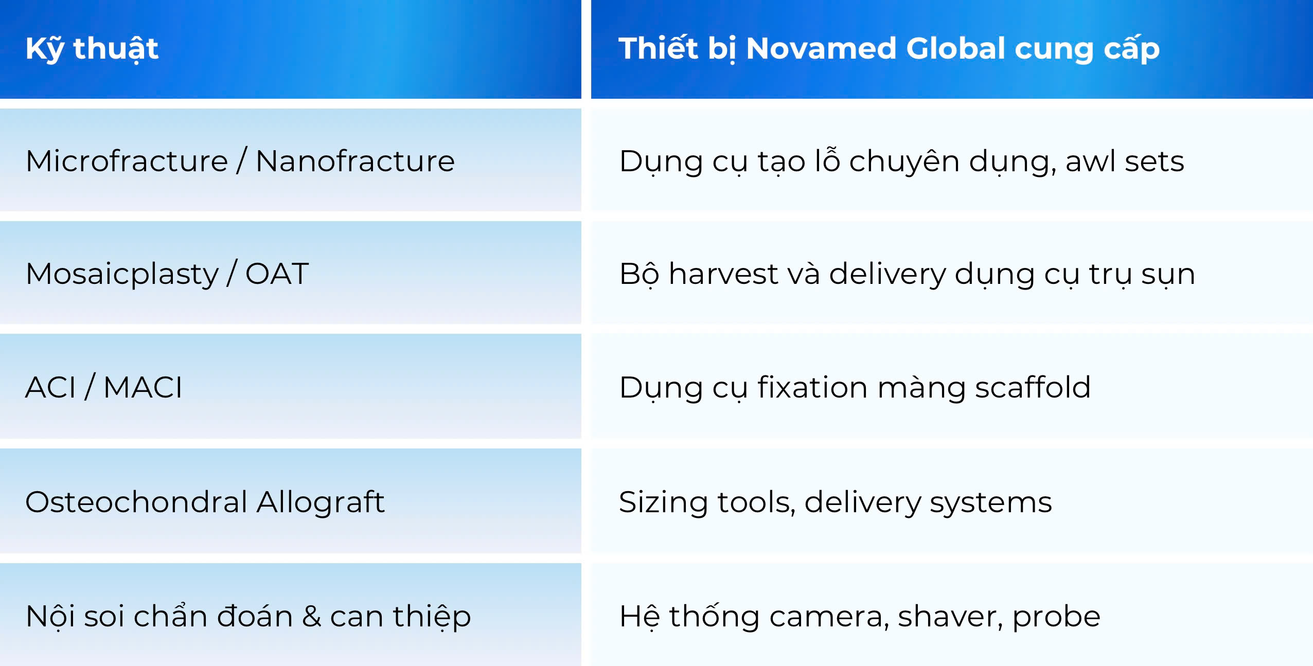
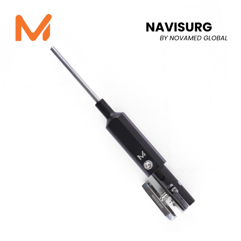
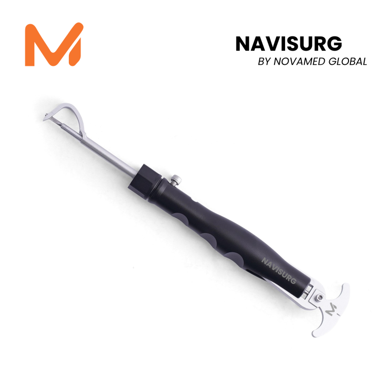
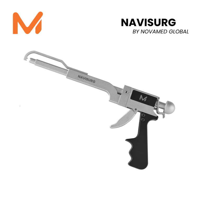
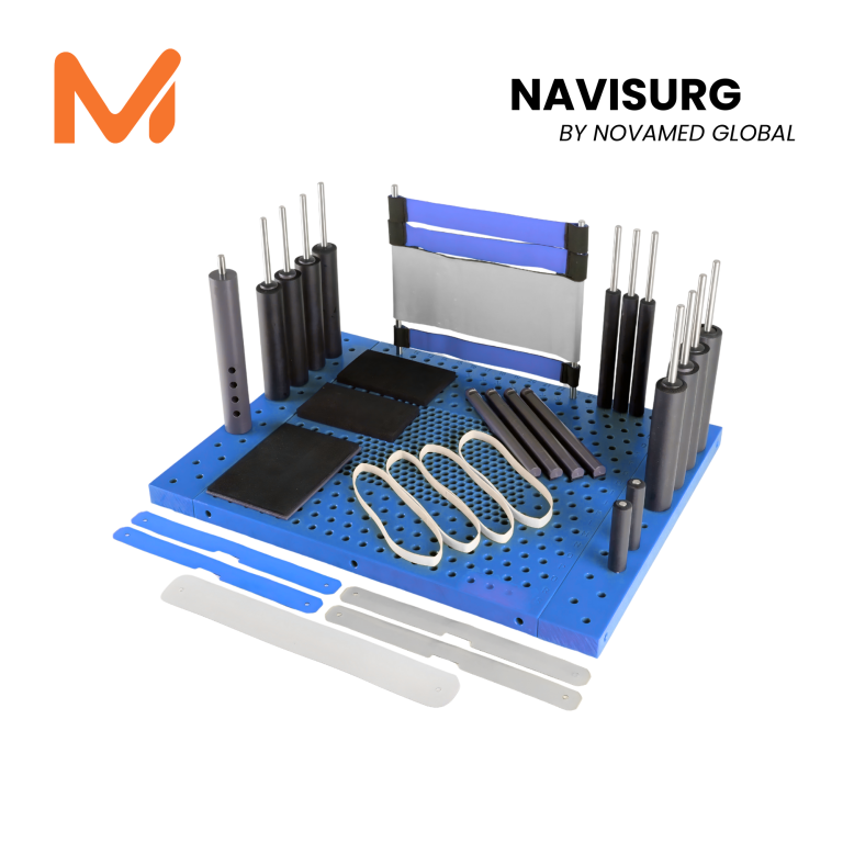
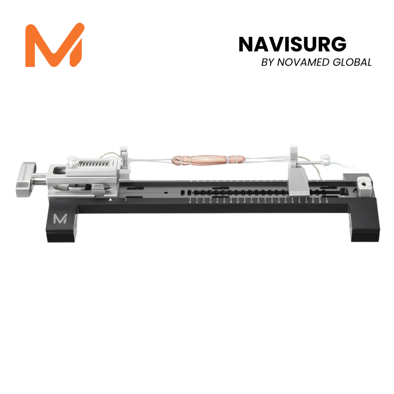
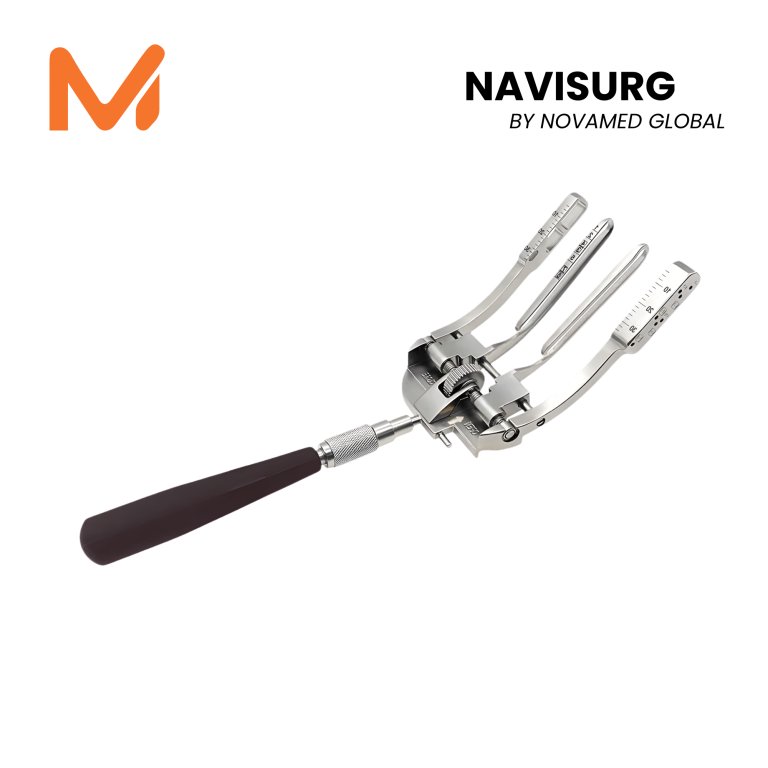
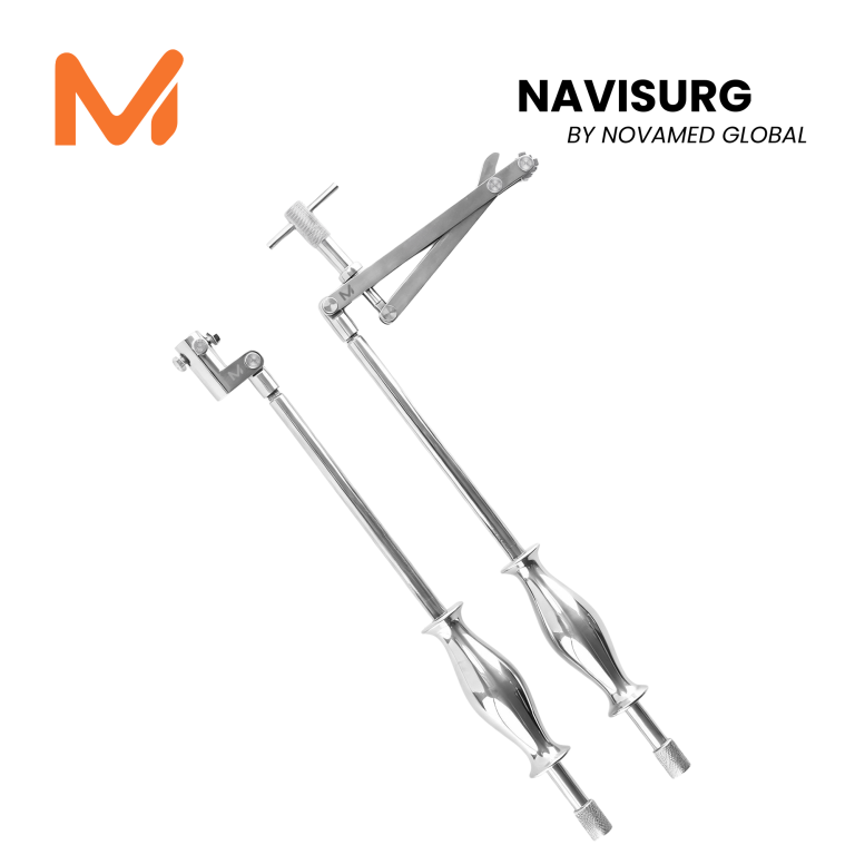
Each cartilage treatment modality requires specialized instruments and implants. Novamed Global provides a comprehensive portfolio of medical devices for healthcare facilities in Vietnam:

Vì Sao Tổn Thương Sụn Khớp Là Thách Thức Đặc Biệt?
Sụn khớp (articular cartilage) là mô đặc biệt bao phủ bề mặt xương trong khớp, giúp khớp trượt êm và phân phối lực tải. Tuy nhiên, đây cũng là một trong những mô khó hồi phục nhất trong cơ thể người.
Sụn khớp gối là mô liên kết hyaline chuyên biệt cao độ, bao gồm tế bào sụn (chondrocytes) và chất nền ngoại bào với 90% là nước cùng proteoglycans, glycosaminoglycans và collagen chủ yếu type II – được tổ chức thành 4 lớp với mật độ tế bào, thành phần chất nền và hướng sợi collagen khác nhau. Đặc điểm mạch máu rất nghèo nàn, tạo ra giới hạn nội tại trong khả năng tự phục hồi của mô này.
Tổn thương sụn khu trú (focal cartilage lesions) là tổn thương hoặc khiếm khuyết khu trú tại bề mặt sụn khớp, thường do chấn thương, mài mòn tích lũy hoặc các bệnh lý khớp tiềm ẩn. Nếu không được điều trị, những tổn thương này có thể gây đau, suy giảm chức năng khớp và góp phần vào sự phát triển của thoái hóa khớp.
Phân Loại Tổn Thương Sụn Khớp Gối
Phân loại theo độ sâu (ICRS Grade)

Phân loại theo tính chất tổn thương
Các chiến lược điều trị được phân thành: giảm nhẹ triệu chứng (chondroplasty, debridement), sửa chữa (drilling, microfracture) và tái tạo (autologous chondrocyte implantation, osteochondral autograft, osteochondral allograft).
Triệu Chứng Và Chẩn Đoán
Lâm sàng
Tổn thương sụn khớp gây ảnh hưởng đến sinh hoạt hằng ngày của bệnh nhân, biểu hiện bằng sưng khớp, hạn chế vận động do đau, đau và căng tức tại chỗ.
Các triệu chứng đặc trưng gồm:
- Đau gối khu trú, tăng khi chịu lực
- Tràn dịch khớp tái phát
- Tiếng kẹt, lạo xạo khi vận động
- Cứng khớp sau nghỉ ngơi dài
- Hạn chế tầm vận động trong trường hợp nặng
Hình ảnh học
- X-quang: Đánh giá chiều cao khe khớp, thoái hóa kèm theo
- MRI: Tiêu chuẩn vàng – xác định vị trí, kích thước, độ sâu tổn thương sụn và tình trạng xương dưới sụn
- Nội soi khớp: Chẩn đoán trực tiếp và can thiệp điều trị đồng thời
Phổ Điều Trị Toàn Diện – Từ Bảo Tồn Đến Tái Tạo
NHÓM 1: Điều Trị Bảo Tồn Và Sinh Học
- Vật lý trị liệu và kiểm soát cân nặng: nền tảng cho mọi phác đồ điều trị, tăng cường cơ quanh khớp, giảm tải trọng lên sụn.
- Tiêm Hyaluronic Acid (Viscosupplementation). Hyaluronic acid cung cấp hiệu quả trong việc giảm triệu chứng đau trong thoái hóa khớp gối. Phù hợp với tổn thương nhẹ-trung bình, đặc biệt là bệnh nhân lớn tuổi chưa đủ điều kiện phẫu thuật.
- PRP (Huyết tương giàu tiểu cầu) cho thấy hiệu quả trong việc giảm triệu chứng và cải thiện chức năng khớp, là phương pháp ít xâm lấn với kết quả hứa hẹn trong việc bảo tồn khớp và sửa chữa sụn.
NHÓM 2: Kỹ Thuật Kích Thích Tủy Xương (Marrow Stimulation)
-
Microfracture – Kỹ Thuật Kinh Điển
Tạo các lỗ nhỏ xuyên qua xương dưới sụn để giải phóng tế bào gốc từ tủy xương, kích thích hình thành mô sụn sợi mới (fibrocartilage).
Các phân tích tổng hợp gần đây chỉ ra rằng kỹ thuật microfracture hiệu quả với các tổn thương nhỏ (đến 4 cm²) trong thời gian theo dõi ngắn hạn. Những hạn chế chính bao gồm: chất lượng sụn sửa chữa kém (fibrocartilage thay vì hyaline), thể tích sụn không ổn định và suy giảm chức năng dài hạn.
Chỉ định tối ưu: Tổn thương < 2–4 cm², bệnh nhân < 40 tuổi, xương dưới sụn còn tốt.
-
Nanofracture và Augmented Microfracture
Thế hệ kế tiếp của microfracture dùng dụng cụ tạo lỗ nhỏ hơn và sâu hơn, kết hợp scaffold sinh học để cải thiện chất lượng mô sửa chữa.
NHÓM 3: Ghép Sụn Xương Tự Thân (Osteochondral Autograft)
Mosaicplasty / OAT (Osteochondral Autograft Transfer)
Lấy trụ sụn-xương từ vùng không chịu lực của khớp gối (thường ở rìa lồi cầu) và ghép vào vị trí tổn thương.
Ưu điểm:
- Ghép sụn hyaline thực sự, chất lượng vượt trội microfracture
- Kết quả ngắn-trung hạn xuất sắc
- Thực hiện một lần phẫu thuật (one-stage)
Giới hạn:
- Phù hợp tổn thương 1-4 cm² (hạn chế nguồn cho)
- Biến chứng vùng cho (donor site morbidity)
Với các tổn thương nhỏ ở bệnh nhân có nhu cầu vận động cao, ghép sụn xương tự thân là lựa chọn điều trị đáng tin cậy.
NHÓM 4: Ghép Sụn Xương Đồng Loại (Osteochondral Allograft – OCA)
Dùng mô sụn-xương từ người hiến tặng để phủ lên vùng tổn thương lớn mà nguồn tự thân không đủ.
Ghép sụn xương tự thân và đồng loại cung cấp giải pháp bền vững với kết quả dài hạn xuất sắc.
Chỉ định: Tổn thương lớn > 4 cm², thất bại với các kỹ thuật trước, bệnh nhân trẻ chưa đủ điều kiện thay khớp.
Lưu ý: Phụ thuộc vào nguồn cung mô, vấn đề bảo quản và nguy cơ truyền bệnh – đòi hỏi quy trình kiểm soát chất lượng nghiêm ngặt.
NHÓM 5: Cấy Sụn Tế Bào Tự Thân – ACI / MACI
Đây là đỉnh cao của tái tạo sụn hiện tại, kỹ thuật phục hồi sụn hyaline thực sự ở người trưởng thành.
Quy trình ACI (2 giai đoạn):
- Giai đoạn 1: Nội soi lấy mẫu sụn nhỏ → nuôi cấy tế bào sụn trong phòng lab (4–6 tuần)
- Giai đoạn 2: Cấy tế bào sụn đã nhân lên vào vị trí tổn thương
MACI (Matrix-induced ACI) – thế hệ tiên tiến hơn: Tế bào sụn được cấy vào màng scaffold collagen type I/III trước khi cấy ghép, tạo thuận lợi cho quá trình tích hợp.
Cấy tế bào sụn tự thân (ACI) đã được sử dụng hơn 20 năm và không ngừng phát triển. Kết quả cho thấy cải thiện kéo dài đến 11 năm với ACI và đến 15 năm với MACI.
Với tổn thương trên 4 cm², ACI vượt trội hơn tất cả các kỹ thuật điều trị khác. Kỹ thuật ACI cho kết quả tốt ở 70-80% bệnh nhân không chỉ trong ngắn hạn mà còn ở theo dõi trung và dài hạn.
NHÓM 6: Scaffold Sinh Học Và AMIC – Hướng Đi Mới
AMIC (Autologous Matrix-Induced Chondrogenesis): Kết hợp microfracture với màng collagen acellular (Chondro-Gide®) – giải pháp một giai đoạn hiệu quả.
Một nghiên cứu RCT cho thấy AMIC là phương pháp điều trị hiệu quả cho tổn thương sụn khu trú, với tác dụng có lợi kéo dài đến 9 năm.
Minced Cartilage (Sụn băm nhuyễn): Kỹ thuật một giai đoạn mới – lấy sụn tự thân, xử lý ngay trong phòng mổ và cấy ghép – đơn giản hóa quy trình so với ACI truyền thống, với kết quả hứa hẹn ở theo dõi 2-5 năm.
Thuật Toán Lựa Chọn Điều Trị Theo Kích Thước Tổn Thương
Kích thước tổn thương sụn

Yếu Tố Quyết Định Kết Quả Điều Trị
Ngoài lựa chọn kỹ thuật, một số yếu tố bệnh nhân ảnh hưởng trực tiếp đến kết quả:
- Tuổi: < 40 tuổi cho kết quả tốt hơn với hầu hết kỹ thuật tái tạo
- BMI: Cao làm tăng tải trọng lên vùng sửa chữa
- Lệch trục chi: Varus/valgus cần được chỉnh trục trước hoặc đồng thời
- Tình trạng xương dưới sụn: Ảnh hưởng lớn đến khả năng tích hợp mảnh ghép
- Thời gian từ chấn thương: Can thiệp sớm cho kết quả tốt hơn
Novamed Global – Giải Pháp Thiết Bị Cho Toàn Bộ Phổ Điều Trị Sụn Khớp
Mỗi kỹ thuật điều trị sụn khớp đòi hỏi thiết bị và dụng cụ chuyên biệt. Novamed Global cung cấp cho các cơ sở y tế Việt Nam danh mục thiết bị toàn diện: