






 Back
BackApplications, News, Education
OSTEOMYELITIS: A DIAGNOSTIC AND THERAPEUTIC APPROACH FROM AN ORTHOPEDIC PERSPECTIVE
Osteomyelitis: A Diagnostic And Therapeutic Approach From An Orthopedic Perspective
Viêm Xương Tuỷ: Tiếp Cận Chẩn Đoán Và Điều Trị Theo Tư Duy Chấn Thương Chỉnh Hình
English Version | Bản tiếng Việt bên dưới.

1. Overview
Osteomyelitis is an infection of bone that induces an acute or chronic inflammatory response, and may be caused by bacteria, fungi, or mycobacteria. Common sources of infection include hematogenous spread, direct inoculation from trauma or open fractures, or postoperative infection associated with implants.
An important point in clinical practice is that osteomyelitis should not be viewed simply as an “infection,” but rather as a condition involving the interaction between infection biology, bone necrosis, and mechanical factors (especially in the presence of implants or unstable fractures).
2. Pathophysiology and the Concepts of “Dead Bone.”
Bone is relatively resistant to infection when intact. However, it becomes susceptible when there is a large initial bacterial inoculum, impaired blood supply or poor tissue perfusion, or the presence of implants that expose surfaces for bacterial adhesion.
2.1. Adhesion – Intracellular Survival – Biofilm
- Staphylococcus aureus has the ability to adhere to extracellular matrix components of bone (laminin, collagen, fibronectin) through adhesins, contributing to persistent infection.
- Bacteria may survive intracellularly within osteoblasts or form biofilms on the surface of bone and orthopedic implants, thereby increasing resistance to treatment and explaining why short antibiotic regimens are associated with higher failure rates.
2.2. Sequestrum – Involucrum – Sinus Tract
- Damage or disruption of the periosteum reduces blood supply to bone, leading to ischemia and necrosis.
- The necrotic bone fragment that separates from viable bone is called a sequestrum. Newly formed bone surrounding the sequestrum is referred to as the involucrum. Drainage from the sequestrum may form a sinus tract.
- In chronic osteomyelitis, necrotic bone is an important histopathological hallmark.
3. Classification: Why Is It Important for Treatment Strategy?
3.1. Acute and Chronic (not solely based on duration)
The classification of acute and chronic osteomyelitis is based more on histopathology than on the duration of disease.
- Acute: before the formation of necrotic bone (usually within the first two weeks, although exceptions may occur).
- Chronic: when necrotic bone has already developed.
3.2. Lew & Waldvogel Classification
This classification is based on the mechanism of infection: hematogenous spread or contiguous spread from adjacent tissues, with or without vascular insufficiency.
3.3. Cierny-Mader Classification (high clinical relevance)
The Cierny-Mader system classifies osteomyelitis according to:
- Anatomical involvement
- Stage 1: medullary
- Stage 2: superficial
- Stage 3: localized
- Stage 4: diffuse
- Host physiological status: A: normal host; B: compromised host – systemic (Bs), local (Bl), or both (Bsl); C: treatment may be worse than the disease.
→ This classification provides the basis for determining the extent of surgical intervention, the need for soft tissue coverage, and the aggressiveness of treatment.
4. Clinical Presentation: When Should Osteomyelitis Be Suspected?
Clinical manifestations depend on the mechanism of infection:
- High-risk situations include bacteremia, endocarditis, injection drug use, and trauma or open fractures.
- Chronic non-healing ulcers in patients with diabetes mellitus, peripheral vascular disease, neuropathy, or the presence of metallic implants should raise suspicion for osteomyelitis.
4.1. Acute
- Onset within approximately two weeks, presenting with localized pain, swelling, warmth, and erythema, sometimes accompanied by fever or chills.
4.2. Subacute
- Patients may present with fatigue and mild pain lasting for several weeks, often without fever.
4.3. Chronic
- Symptoms are prolonged, and fever may be absent; recurrent swelling, pain, and the presence of sinus tracts may occur.
4.4. Special form: Native Vertebral Osteomyelitis (NVO)
- New or worsening back or neck pain accompanied by fever and elevated inflammatory markers, particularly in the presence of bacteremia or endocarditis, should raise suspicion for NVO.
5. Physical Examination: Key Points in Patients with Foot Ulcers
- Clinical examination should focus on identifying the source of infection (nidus), as well as assessing sensation and peripheral vascular status.
- Probe-to-bone test: using a sterile blunt instrument to probe the ulcer. If bone can be contacted through the ulcer, this strongly suggests osteomyelitis (particularly in diabetic foot ulcers) and helps determine whether imaging studies or biopsy are required.
6. Laboratory and Imaging Studies
6.1. Blood Tests
- White blood cell count may be normal, while ESR and CRP are often elevated but nonspecific.
- CRP is useful for monitoring treatment response.
- Blood cultures may be positive, particularly in hematogenous osteomyelitis (for example, in NVO).
6.2. Imaging: Choosing the Appropriate Modality
Plain radiography:
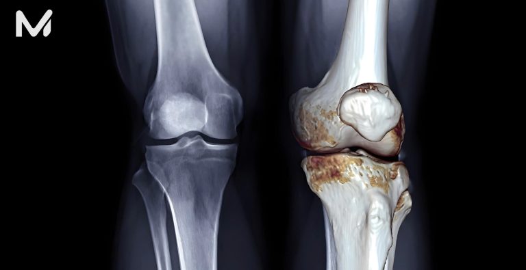
- Usually, the initial imaging modality. However, radiographic changes typically appear only after approximately 14 days, when 50–75% of bone mineral content has been lost, making it less useful in early disease.
- Suggestive findings include soft tissue swelling, osteopenia, osteolysis, bone destruction, and periosteal reaction.
MRI:
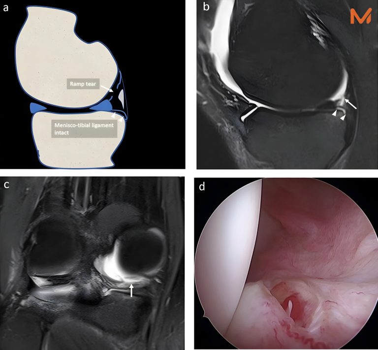
- Highly sensitive for evaluating the extent of infection, bone marrow involvement, abscess formation, and necrosis.
Bone scintigraphy with Tc-99m or labeled leukocyte scans:
- Highly sensitive but less specific; useful when an MRI cannot be performed due to metallic implants.
Contrast-enhanced CT:
- Does not necessarily improve the detection of disease itself, but may help differentiate phlegmon, necrotic tissue, and abscess in certain situations.
7. Principles of Definitive Diagnosis: “Microbiology from Bone or Deep Tissue.”
In clinical practice, the primary goal is to identify the causative pathogen in order to guide targeted antimicrobial therapy.
Clinical consideration: Obtaining microbiological samples before initiating antibiotic therapy, when feasible, significantly increases the likelihood of identifying the pathogen.
8. Treatment: Antibiotics and Surgery Based on Bone Biology
8.1. Why Is Surgery Often Necessary?
Antibiotics penetrate poorly into abscess cavities, necrotic tissue, and dead bone; therefore, debridement of necrotic bone and soft tissue is often required.
8.2. When Implants or Hardware Are Present
In osteomyelitis associated with prosthetic joints or orthopedic implants, hardware removal is usually recommended.
However, in selected cases where the joint remains stable (for example, the hip joint) and the pathogen is highly susceptible (such as streptococci), prolonged antibiotic therapy for several months without hardware removal may be considered.
8.3. Treatment Strategy Framework (Suggested for Implementation in NOVAMED)
Pillar 1 – Surgical control of infection
- Adequate surgical exposure and thorough debridement of necrotic tissue and abscess.
- Management of dead space.
- Mechanical stabilization when fractures or bone defects are present (important for bone healing).
Pillar 2 – Antibiotic therapy
- Antibiotics should be selected based on the identified pathogen and susceptibility profile. CRP may be used as a marker of treatment response.
- Treatment duration depends on disease type (acute or chronic), the presence of hardware, treatment response, and the extent of debridement.
Pillar 3 – Optimization of host factors
- Control of diabetes mellitus, nutritional status, vascular disease, and smoking cessation.
- For foot ulcers: vascular assessment, off-loading, and appropriate wound care are essential.
9. Important Differential Diagnoses (to Avoid Overdiagnosis)
The following conditions should be considered in the differential diagnosis, particularly in patients with diabetes:
- Charcot arthropathy
- SAPHO syndrome
- Rheumatoid arthritis
- Bone metastasis
- Fractures (including stress or pathologic fractures)
- Gout
- Avascular necrosis
10. Prognosis and Follow-up
- Acute osteomyelitis generally has a favorable prognosis if diagnosed and treated early.
- Chronic osteomyelitis may recur, particularly in patients with impaired immunity or new trauma to the previously affected area.
- Patient education regarding the prolonged duration of treatment and adherence to therapy is crucial to reduce recurrence.
- A multidisciplinary approach is essential, involving orthopedic surgeons, radiologists, infectious disease specialists, wound care teams, vascular specialists, and rehabilitation physicians.
1. Tổng quan
Viêm xương tủy là tình trạng nhiễm trùng xương gây phản ứng viêm cấp hoặc mạn, có thể do vi khuẩn, nấm hoặc mycobacteria. Nguồn lây thường gặp gồm đường máu, trực tiếp do chấn thương/gãy hở, hoặc sau phẫu thuật implant.
Điểm cần nhấn mạnh trong thực hành: đây không chỉ là “nhiễm trùng”, mà là một bệnh lý kết hợp giữa sinh học nhiễm trùng – hoại tử xương – và cơ học (đặc biệt khi có implant hoặc ổ gãy không vững).
2. Cơ chế bệnh sinh và các khái niệm “xương chết”
Xương là mô tương đối “kháng nhiễm” khi còn nguyên vẹn; xương trở nên dễ nhiễm khi có lượng vi khuẩn xâm nhập ban đầu lớn, thiếu máu, thiếu nuôi dưỡng, hoặc implant làm lộ bề mặt cho vi khuẩn bám dính.
2.1. Bám dính – Sống nội bào – Biofilm
- S. aureus có khả năng bám vào thành phần nền xương (laminin, collagen, fibronectin…) thông qua các adhesins, góp phần làm nhiễm trùng dai dẳng.
- Vi khuẩn có thể tồn tại nội bào trong osteoblast hoặc tạo biofilm trên bề mặt xương và dụng cụ → Tăng “kháng” điều trị, giải thích vì sao các phác đồ quá ngắn có tỷ lệ thất bại cao.
2.2. Sequestrum – Involucrum – Sinus tract
- Tổn thương hoặc gián đoạn màng xương làm giảm tưới máu → Thiếu máu xương → Hoại tử.
- Phần xương hoại tử tách rời gọi là sequestrum, xương mới hình thành bao quanh gọi là involucrum . Dịch thoát ra từ sequestrum có thể tạo đường rò (sinus tract).
- Trong viêm xương tủy mạn, xương hoại tử là dấu ấn mô bệnh học quan trọng.
3. Phân loại: Vì sao quan trọng đối với chiến lược điều trị?
3.1. Cấp và mạn (không chỉ dựa vào thời gian)
Phân loại cấp và mạn dựa trên mô bệnh học hơn là “số tuần mắc bệnh”:
- Cấp: trước khi hình thành xương hoại tử (thường trong khoảng 2 tuần đầu, nhưng có ngoại lệ)
- Mạn: khi đã có xương hoại tử
3.2. Lew & Waldvogel
Phân loại theo cơ chế: đường máu và lan liên tục (contiguous) và có hoặc không tình trạng thiếu máu nuôi (vascular insufficiency).
3.3. Cierny-Mader (giá trị thực hành cao)
Cierny-Mader phân loại theo:
- Giải phẫu tổn thương:
- Stage 1: tủy xương
- Stage 2: nông
- Stage 3: khu trú
- Stage 4: lan tỏa
- Tình trạng cơ địa bệnh nhân: A (bình thường), B (có yếu tố suy giảm toàn thân – Bs / tại chỗ – Bl / cả hai – Bsl), C (điều trị “tệ hơn bệnh”).
→ Đây là nền tảng để quyết định mức độ phẫu thuật, nhu cầu che phủ mô mềm và “độ quyết liệt” của điều trị.
4. Lâm sàng: Khi nào cần nghĩ đến viêm xương tủy?
Biểu hiện phụ thuộc cơ chế nhiễm:
- Nguy cơ cao: nhiễm khuẩn huyết (bacteremia), viêm nội mạc (endocarditis), tiêm chích, chấn thương hoặc gãy hở.
- Loét mạn khó lành ở bệnh nhân đái tháo đường, bệnh mạch máu ngoại biên, bệnh lý thần kinh (neuropathy), có dụng cụ kim loại… phải nghĩ đến viêm xương tủy.
4.1. Cấp tính
- Khởi phát trong khoảng 2 tuần: đau, sưng nóng đỏ tại chỗ, có thể sốt hoặc ớn lạnh.
4.2. Bán cấp
- Mệt mỏi, đau nhẹ kéo dài vài tuần, ít sốt.
4.3. Mạn tính
- Triệu chứng kéo dài, sốt có thể không rõ; có thể có đường rò, sưng đau tái phát.
4.4. Đặc biệt: viêm xương cột sống (Native Vertebral Osteomyelitis – NVO)
- Đau lưng, cổ mới xuất hiện hoặc nặng lên kèm sốt, triệu chứng viêm tăng, nhất là có bacteremia/endocarditis → Cần nghĩ NVO.
5. Thăm khám: “điểm mấu chốt” ở bệnh nhân loét bàn chân
Thăm khám nên tập trung:
- Tìm “ổ nguồn” (nidus), đánh giá cảm giác, mạch máu ngoại biên.
- Probe-to-bone test: dùng dụng cụ vô khuẩn đầu tù chạm được xương qua ổ loét là dấu hiệu gợi ý mạnh viêm xương tủy (đặc biệt trong bàn chân ĐTĐ), giúp quyết định có cần chẩn đoán hình ảnh hoặc sinh .
6. Cận lâm sàng và hình ảnh
6.1. Xét nghiệm máu
- WBC có thể bình thường; ESR/CRP tăng không đặc hiệu.
- CRP hữu ích để theo dõi đáp ứng điều trị.
- Cấy máu có thể cho kết quả dương tính, đặc biệt trong viêm xương tủy đường máu (ví dụ: NVO).
6.2. Hình ảnh học: chọn đúng “bậc thang”
X-quang thường quy:
- Thường là bước đầu, nhưng có độ trễ khoảng 14 ngày mới thấy dấu hiệu; phát hiện tổn thương tiêu xương khi đã mất 50-75% nền xương → kém cho giai đoạn sớm.
Dấu gợi ý:
- Sưng phần mềm, osteopenia, osteolysis, phá hủy xương, phản ứng màng xương.
MRI:
- Nhạy trong đánh giá phạm vi viêm, tủy xương, ổ áp-xe/hoại tử (MRI là một trong các xét nghiệm hình ảnh hữu ích nhất trong đánh giá).
Xạ hình xương Tc-99m / Xạ hình bạch cầu đánh dấu:
- Nhạy cao nhưng đặc hiệu kém, hữu ích khi không chụp MRI được do dụng cụ kim loại.
CT có thuốc cản quang:
- Không làm tăng phát hiện bệnh bằng contrast chính, nhưng giúp phân biệt phlegmon – mô hoại tử – áp-xe trong một số tình huống.
7. Nguyên tắc chẩn đoán xác định: “vi sinh từ xương/mô sâu”
Trong thực hành, mục tiêu là xác định tác nhân để điều trị đích.
Lưu ý lâm sàng: Lấy mẫu trước khi kháng sinh, khi tình trạng bệnh nhân cho phép, thường giúp tăng khả năng tìm tác nhân.
8. Điều trị: kháng sinh và phẫu thuật theo sinh học của xương
8.1. Vì sao phẫu thuật thường cần thiết?
Kháng sinh thấm kém vào ổ áp-xe, mô hoại tử và xương chết; vì vậy, debridement xương/mô hoại tử thường là chỉ định khi có thể.
8.2. Khi có implant hoặc hardware
Trong viêm xương liên quan đến khớp giả hoặc implant, tháo dụng cụ thường được chỉ định.
Tuy nhiên, cũng có trường hợp ngoại lệ: nếu khớp ổn định (ví dụ khớp háng) và tác nhân nhạy cao (như streptococci), có thể cân nhắc điều trị kháng sinh kéo dài nhiều tháng mà không tháo (chọn lọc).
8.3. Khung chiến lược điều trị (gợi ý triển khai trong NOVAMED)
Trụ cột 1 – Kiểm soát ổ nhiễm bằng phẫu thuật
- Mở đường vào đủ rộng, cắt lọc triệt để mô hoại tử/áp-xe
- Xử trí khoang chết
- Ổn định cơ học nếu có gãy hoặc khuyết xương (quan trọng để liền xương)
Trụ cột 2 – Kháng sinh
- Dựa trên tác nhân và độ nhạy, theo dõi CRP như marker đáp ứng.
- Thời gian điều trị phụ thuộc vào thể bệnh (cấp và mạn, có hardware, đáp ứng, mức độ debridement).
Trụ cột 3 – Tối ưu cơ địa bệnh nhân
- Kiểm soát đái tháo đường, dinh dưỡng, bệnh mạch máu, cai thuốc lá…
- Với loét bàn chân: đánh giá mạch máu, off-loading, chăm sóc vết thương chuẩn.
9. Chẩn đoán phân biệt cần nhớ (tránh chẩn đoán quá mức)
Sau đây là liệt kê các chẩn đoán phân biệt quan trọng, đặc biệt ở bệnh nhân đái tháo đường:
- Charcot arthropathy
- SAPHO
- Viêm khớp dạng thấp
- Di căn xương
- Gãy xương (kể cả stress/pathologic)
- Gout
- Hoại tử vô mạch
10. Tiên lượng và theo dõi
- Viêm xương cấp, nếu điều trị sớm, thường tiên lượng tốt.
- Viêm xương mạn có thể tái phát, nhất là khi miễn dịch suy giảm hoặc có chấn thương mới tại vùng đã điều trị.
- Giáo dục bệnh nhân về thời gian điều trị kéo dài và tuân thủ là yếu tố then chốt để giảm tái phát.
- Cách tiếp cận đa chuyên khoa: ngoại chấn thương chỉnh hình, chẩn đoán hình ảnh, truyền nhiễm, chăm sóc vết thương, mạch máu, phục hồi chức năng…